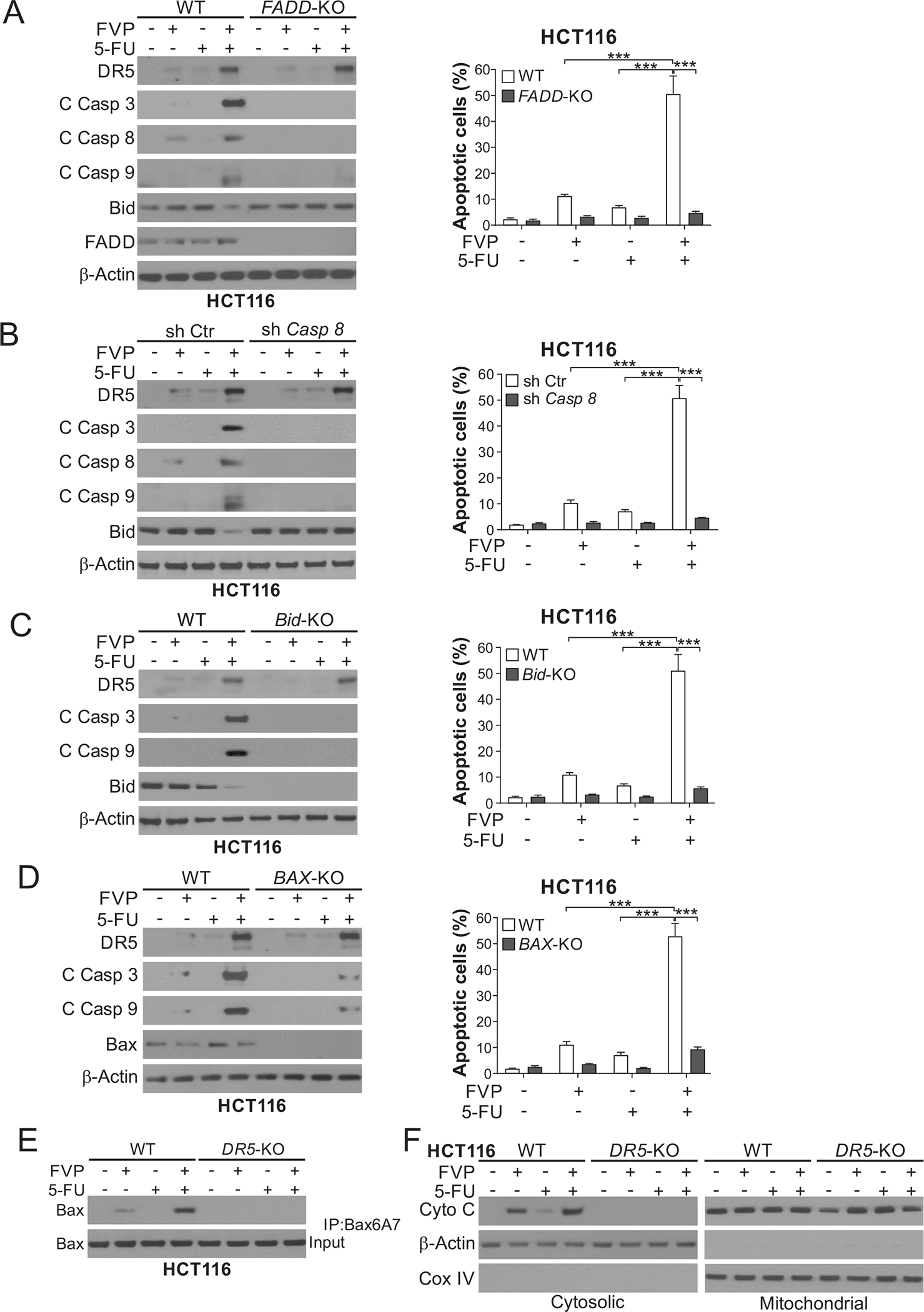
Figure 5. DR5 mediates apoptosis induced by CDKIs combined with 5-FU via crosstalk of the intrinsic and extrinsic pathways.

(A)-(D) WT HCT116 cells along with isogenic (A) FADD-KO, (B) stable caspase 8 shRNA knockdown (Casp 8-KD) and shRNA control (sh Ctr), (C) Bid-KO, and (D) BAX-KO HCT116 cells were treated with 20 nM FVP, 15 μg/mL 5-FU, or their combination for 24 hours. Left panels: Western blotting of indicated proteins; right panels: analysis of apoptosis by counting cells with condensed and fragmented nuclei after nuclear staining with Hoechst 33258. (E), (F) WT and DR5-KO HCT116 cells were treated as in (A)-(D). (E) Bax conformational change was analyzed by immunoprecipitation (IP) with anti-Bax 6A7 (activated) antibody followed by Western blotting. (F) Cytochrome c release was analyzed by Western blotting of mitochondrial and cytosolic fractions isolated from treated cells. β-Actin and cytochrome oxidase subunit IV (COX IV) were used as controls for loading and fractionation. In (A)-(D), values were expressed as means ± SD of three independent experiments. ***, P < 0.001.
