
Figure 4.
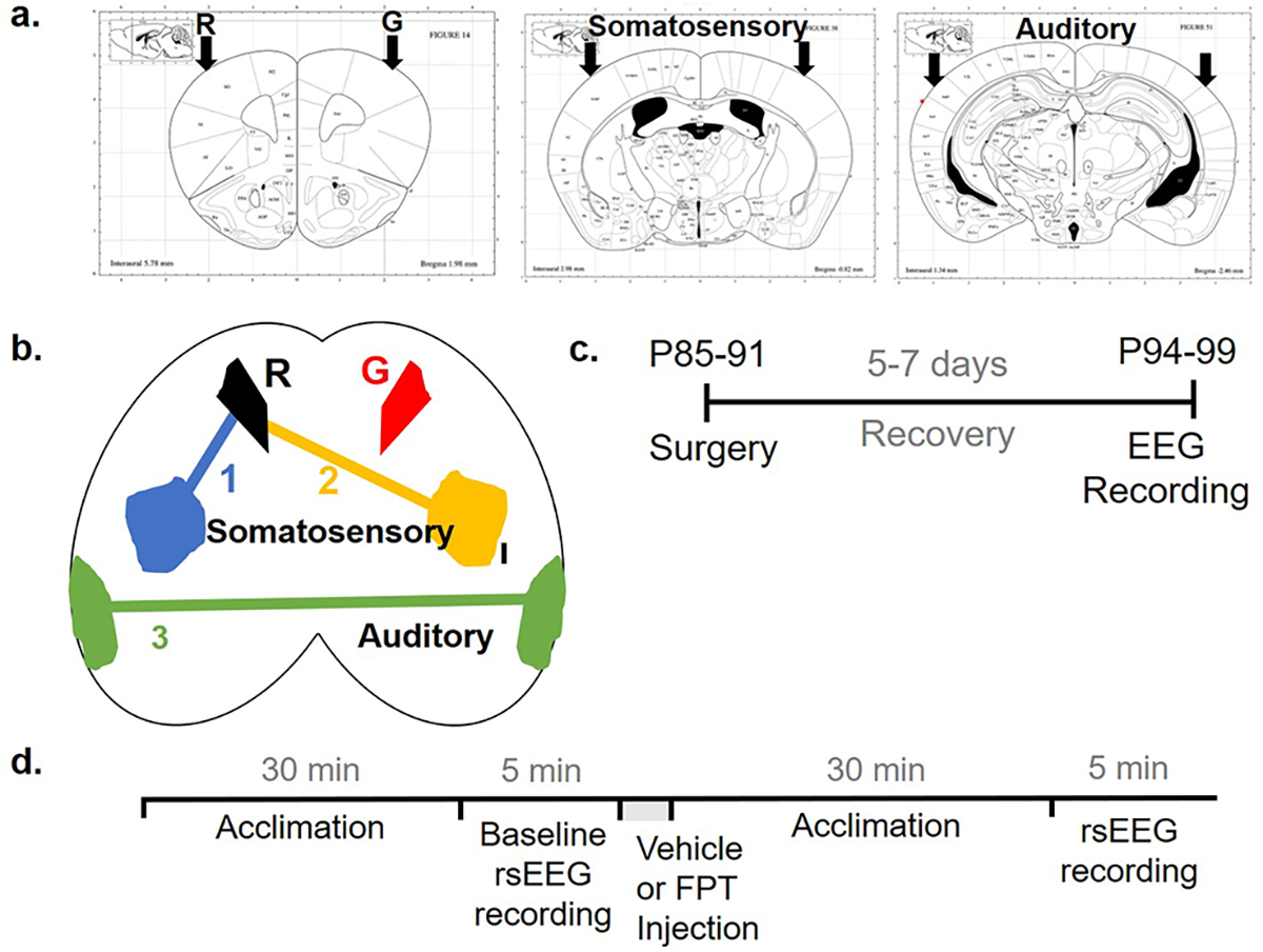
EEG surgery coordinates and timelines. Electrode coordinates and channels are shown in coronal (a) and axial (b) planes. Timeline for surgery (c) and recording (d) of non-task-related, resting-state (rs) EEG. R, reference electrode; G, ground electrode.