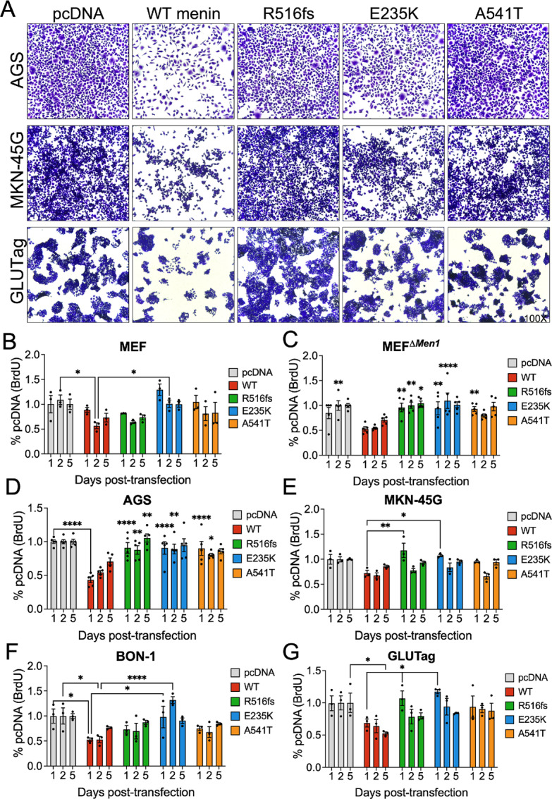
FIGURE 5.

Clinical MEN1 mutations and variants exhibit loss of growth-suppressive function in a cell-dependent manner. A, Representative bright-field images of crystal violet-stained AGS, MKN-45G, and GLUTag following 72 hours transfection with empty vector (pcDNA), wild-type (WT) menin, and the three mutated menin proteins. Images taken at 100X magnification. BrdU incorporation was assayed 1, 2, and 5 days posttransfection in MEF (B), MEFΔMen1 (C), AGS (D), MKN-45G (E), BON-1 (F), and GLUTag (G) cell lines. n = 3–5 experimental replicates. Asterisks represent comparisons between each timepoint. *, P < 0.05; **, P < 0.01; ****, P < 0.0001 by two-way ANOVA with Tukey post hoc test; mean ± SEM.
