
FIGURE 3.
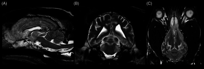
Sagittal (A), transverse (B), and dorsal (C) T2W sequence showing regression of the hydrocephalus compared to the state before the shunt placement. Images displayed in radiological convention: right side of the dog is to the left of the image, rostral is to the left of the image.