
FIGURE 3.
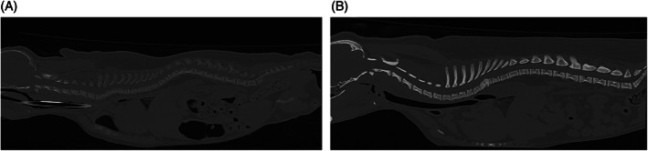
(A) Computed tomography images of the entire spine of Pug 1.1♀ with VDDR type 1A showing generalized decrease in bone density (attenuation). Endplate widening is also noted. (B) Computed tomography images of the entire spine of an age‐matched control pug.