
FIGURE 4.
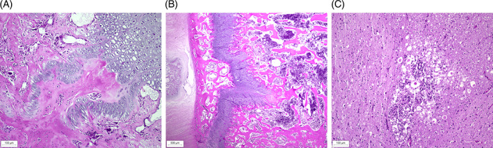
(A) Histopathological changes at sites of enchondral ossification in the bones of Pug 1.1♀ with VDDR type 1A. Costochondral joint, longitudinal section stained with hematoxylin and eosin (H&E), showing retention of hypertrophic chondrocytes and tongue‐like projections of cartilage extending into the metaphysis from the physeal cartilage. (B) Histopathological changes at sites of enchondral ossification in the bones of Pug 1.1♀ with VDDR type 1A. Vertebrae T13–L1, transverse section (H&E), showing disorganized columns of hypertrophic chondrocytes and tongue‐like projections of cartilage in the metaphysis. (C) Spinal cord at T13–L1, transverse section (H&E), showing focal malacia with moderate parenchymal destruction and gliosis of primarily the ventral funiculi at the level of spinal cord stenosis.