
FIGURE 3.
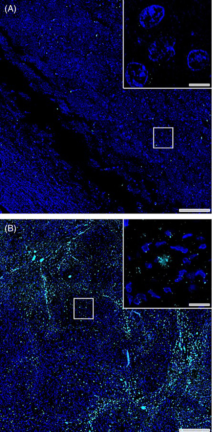
Immunofluorescence staining of tyrosine hydroxylase‐rich crystals. Immunofluorescence staining of tyrosine hydroxylase (cyan) and cellular nuclei (DAPI, blue) in tissue with unremarkable brain pathology (A) and within biopsy tissue section of case study patient (B). Scale bars in low magnification montage images = 200 μM and scale bars in high magnification insets = 10 μM.