
FIGURE 3.
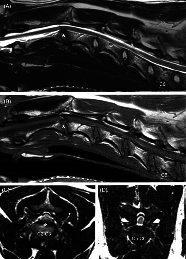
Sagittal T2 (A) and T1‐weighted (B) and transverse T2‐weighted (C, D) MR images from a 6‐year‐old Great Dane. Compression of the spinal cord can be seen at C2‐C3 because of intervertebral disc protrusion (asterisk), and at C5‐C6 caused by osseous proliferation of the articular processes (D—arrows). There is reduced articular joint fluid bilaterally at C5‐C6 (D). Note the presence of an artifact on sagittal T2w and T1W images over the spinal cord at C2‐C3.