
FIGURE 3.
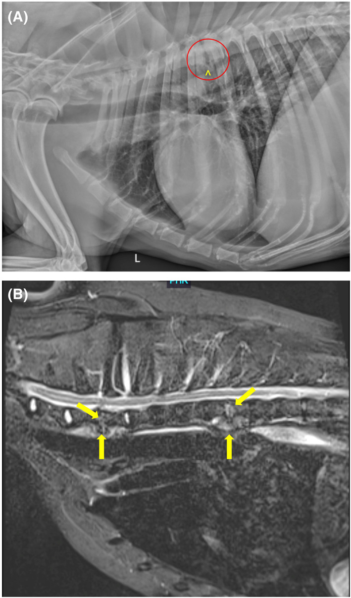
(A and B) Representative images of a 9‐year‐old male neutered Labrador Retriever with discospondylitis. Advanced imaging such as MRI may detect more lesions than are evident on routine radiographs. (A) A left lateral radiographic projection reveals lysis of the caudal endplate of T5 and the cranial endplate of T6 (within red circle), along with spondylosis deformans at T5‐T6 (^). (B) A midline STIR image of the cranial thoracic spine reveals irregularity and hyperintensity of the C7‐T1 and T5‐T6 intervertebral discs and adjacent cranial and caudal vertebral endplates (arrows).