
FIGURE 3.
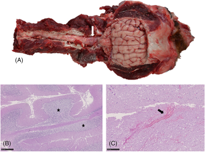
Gross pathology and histopathology of the brain of a 2.5‐year‐old intact male spaniel crossbreed dog that was euthanized after in‐hospital cardiopulmonary arrest. (A) Head and proximal cervical spinal cord with the caudodorsal calvarium and dorsal vertebral column removed. The cerebellum is positioned caudally within the skull (associated with in situ foramen magnum herniation) and the brainstem and proximal cervical spinal cord are reddened (arrow). (B) Cerebellum, Hematoxylin & eosin (H&E). Mild cerebellar edema in the granular cell layer (asterisk). Scale bar, 500 μm. (C) Proximal cervical spinal cord, H&E. Multifocal areas of acute hemorrhage expanding the gray and white matter (arrow). Scale bar, 250 μm.