
FIGURE 4.
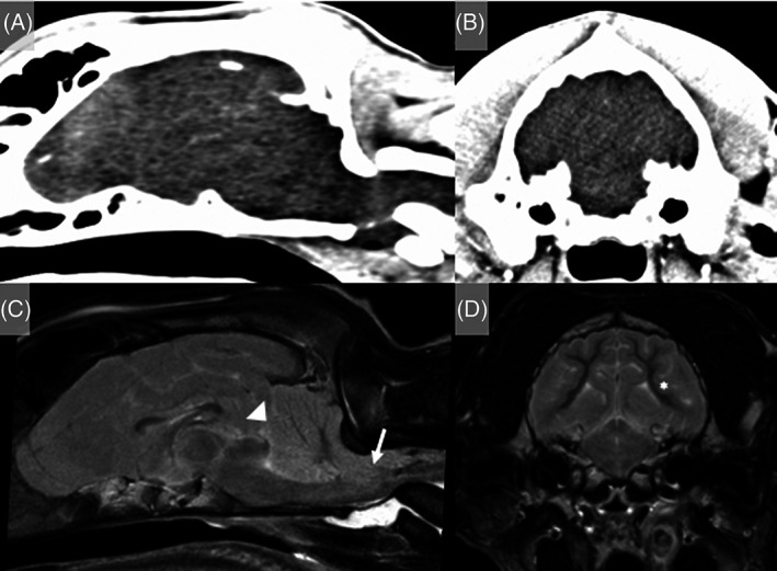
A 6‐month‐old intact male Weimaraner that presented after an out‐of‐hospital cardiopulmonary arrest secondary to trauma. (A and B) CT reconstruction of the head showed no evidence of head trauma. (C and D) MRI of the head (T2W sagittal [C] and transverse image at the level of the rostral colliculi [D]) identified diffuse loss of differentiation between cerebral gray and white matter, diffuse cortical hyperintensity on T2W with swelling of the cortical gray matter (asterisk), loss of cerebral sulci, and severe foramen magnum (arrow) and caudal transtentorial herniation (arrowhead) with secondary brainstem compression. These changes were consistent with a global HIBI and euthanasia was elected given the severity of the clinical presentation and MRI findings. Necropsy was not performed. Hyperintensities are reported relative to normal gray matter.