
FIGURE 5.
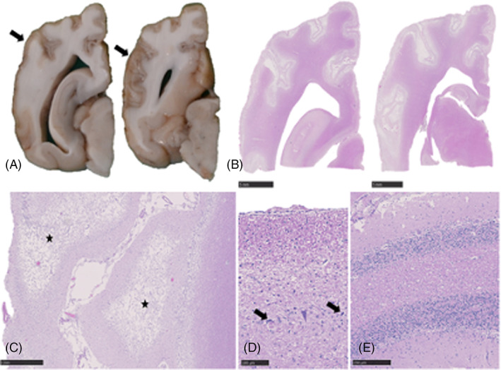
Gross pathology and histopathology of the brain of a 7‐month‐old intact male Cocker Spaniel with asphyxiation secondary to laryngeal obstruction. (A) Cerebrum, formalin fixed serial sections. Severe thinning and discoloration of the parietal and temporal cerebral cortex (arrows). (B) Cerebrum, H&E sections corresponding to (A). Diffuse pallor of the cortical gray matter with areas of neuropil loss. Scale bars, 5 mm. (C) Occipital cortex, H&E. Severe cortical spongiosis and necrosis (asterisk) with neovascularization. Scale bar, 1 mm. (D) Hippocampus, CA1 region, H&E. Severe neuronal degeneration (arrow) and loss. Scale bar, 100 μm. (E) Cerebellar folia, H&E. Marked Purkinje cell loss. Scale bar, 250 μm.