
FIGURE 3.
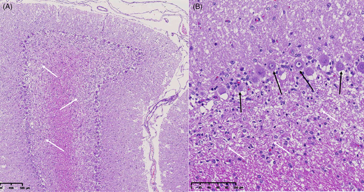
Histopathology of the cerebellum of a dog with granuloprival degeneration showing thinning of the granule cell layer because of a massive loss of granule cells (white arrows), with relative sparing of Purkinje cells (black arrows). Courtesy of Martí Pumarola Battle.