
FIGURE 6.
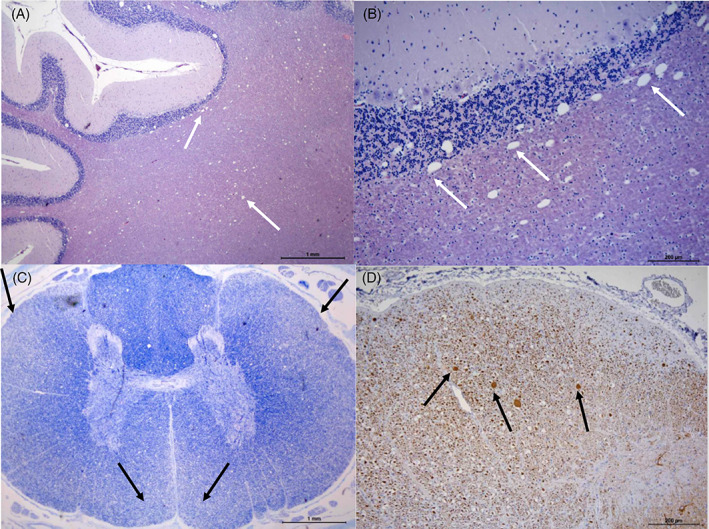
Histopathology of the spinal cord and cerebellum of a Bouvier des Ardennes dog affected with SAMS. (A,B) HE stain shows spongiosis (spongy degeneration) in the cortical white matter and infiltrating the cortical cerebellar granular layer (white arrows). (C) Luxol fast blue (LFB) stain of the spinal cord shows marked pallor of lateral and ventral funiculi (black arrows). (D) IHC of dorsal spinocerebellar tract of cervical spinal cord showing decreased immune positivity for NF200, loss of axonal component and presence of some spheroids (black arrows). Courtesy of Martí Pumarola Battle.