
FIGURE 9.
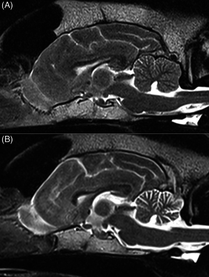
Sagittal T2W MRI image (1.5T) of the brain of a normal dog (A) and of an American Staffordshire Terrier with neuronal ceroid lipofuscinosis 4A, showing marked diffuse atrophy of the cerebellar cortex (B). Courtesy of Ariel Cohen Solal.
Official websites use .gov
A
.gov website belongs to an official
government organization in the United States.
Secure .gov websites use HTTPS
A lock (
) or https:// means you've safely
connected to the .gov website. Share sensitive
information only on official, secure websites.
Sagittal T2W MRI image (1.5T) of the brain of a normal dog (A) and of an American Staffordshire Terrier with neuronal ceroid lipofuscinosis 4A, showing marked diffuse atrophy of the cerebellar cortex (B). Courtesy of Ariel Cohen Solal.