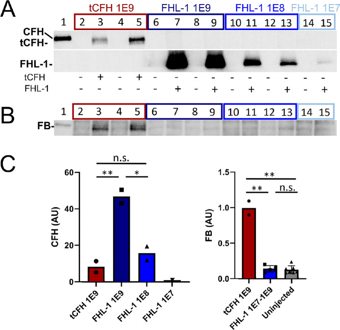
Figure 3.
Posterior eyecup expression of endogenous FB in Cfh–/– mice is CFH construct dependent. CFH (A) and FB (B) immunoblots of eyecup (RPE/BrM/choroid/sclera) lysates isolated from Cfh–/– mice following subretinal injections with AAVs expressing tCFH (lanes 3 and 5, red box) or FHL-1 (lanes 7, 9, 11, 13, and 15; dark blue, blue, and light blue boxes). Corresponding dose vector genomes (vg) of each subretinal injection are shown on the x-axis of the graphs in C. The non-injected control contralateral eyecup lysates (–) were loaded in the even-numbered lane to the left of each injected eyecup lysate (+). Lane 1 is a positive control for full-length CFH (CFH H/H mouse plasma). (C) Densitometric analysis of immunoblots in A and B. The relative protein levels measured by densitometry are depicted in the bar graphs (CFH, left panel; FB, right panel). The CFH values are normalized to the single eye with FHL-1 from the lowest dose group (1E7 vg, lane 15, light blue box). Even though the levels of FHL-1 are about sixfold higher than tCFH at an equivalent viral dose, only the two eyecup lysates from eyes expressing the tCFH have intact FB. *P < 0.05; **P < 0.01.

