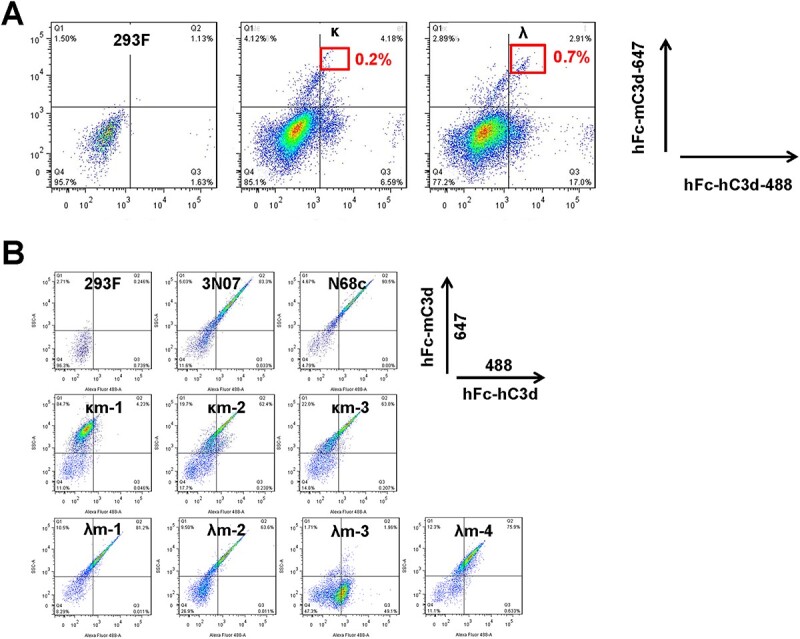
Figure 6.

Enrichment and screen of cells displaying antibodies with cross-reactivity. (A) The top 0.2% and top 0.7% double-positive cells from both sublibraries were sorted to obtain single cells after staining by hFc-hC3d conjugated to Alexa Fluor 488 and hFc-mC3d conjugated to Alexa Fluor 647 in presence of 10% PNHCS. (B) Single-cell clones with binding activity to both hC3d and mC3d were verified by dual staining with hFc-hC3d conjugated to Alexa Fluor 488 and hFc-mC3d conjugated to Alexa Fluor 647 in presence of 10% PNHCS.
