
Fig. 3.
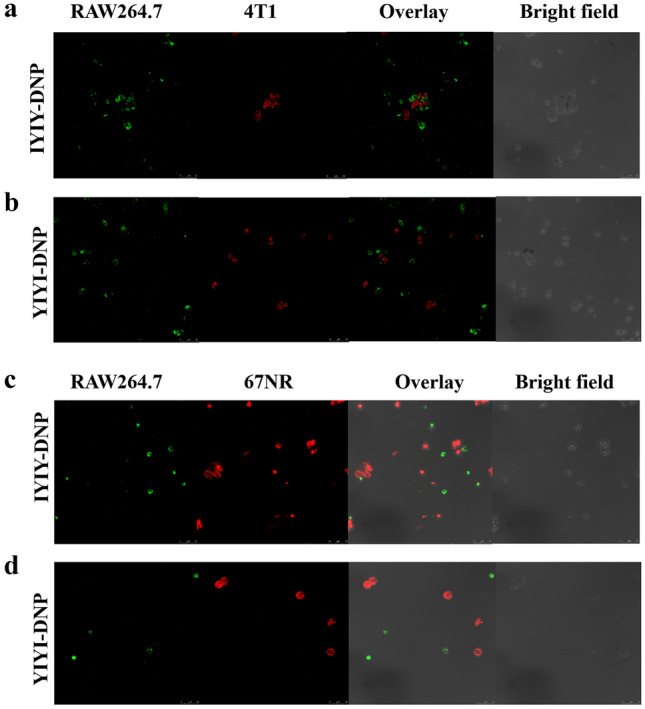
In vitro antibody-dependent cellular phagocytosis in the presence of 0.1% of anti-DNP antibodies. RAW264.7 macrophages were labelled with green dye whereas cancer cells (4T1, 67NR) were labelled with red dye. a 4T1 with 10 µM IYIY-DNP, b 4T1 with 10 µM YIYI-DNP, c 67NR with 10 µM IYIY-DNP, d 67NR with 10 µM YIYI-DNP. Data shown are representative of at least five fields, three independent experiments (n = 3) with similar results