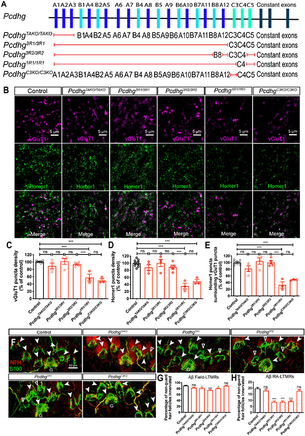
Figure 5. Differential requirements of Pcdhg isoforms for synapse formation and peripheral axonal branching.
(A) Summary of the Pcdhg mutants used. Red lines indicate the corresponding genes are disrupted, while the genes listed are not disrupted.
(B) IHC images of spinal cord dorsal horn lamina III from P21 control and Pcdhg mutants.
(C-E) Normalized densities of vGluT1+ (C) and Homer1+ (D) puncta from P21 control and various Pcdhg mutants. (E) shows the normalized densities of Homer1+ puncta that surrounds vGluT1+ puncta. One-way ANOVA with Tukey’s post hoc test. Each dot represents average value of an animal.
(F) Whole-mount immunostaining of adult back hairy skin sections from control and various Pcdhg mutants. Aβ field-LTMRs innervations are noted by white arrowheads. Hair follicles without any NFH+ Aβ field-LTMRs are denoted by empty arrowheads. Scale bar represents 50 μm.
(G and H) Quantification of the percentage of non-guard hair follicles innervated by NFH+ Aβ field-LTMRs circumferential endings (G) and Aβ RA-LTMRs lanceolate endings (H). n = 649 hair follicles from 5 wildtype animals; n = 336 hair follicles from 2 PcdhgTAKO animals; n = 710 hair follicles from 4 Pcdhg3R1 animals; n = 673 hair follicles from 5 Pcdhg3R2 animals; n = 614 hair follicles from 3 PcdhgC3KO animals. One-way ANOVA with Tukey’s post hoc test.
ns, not significant; *p < 0.05; **p < 0.01; ***p < 0.001.

