
Figure 3.
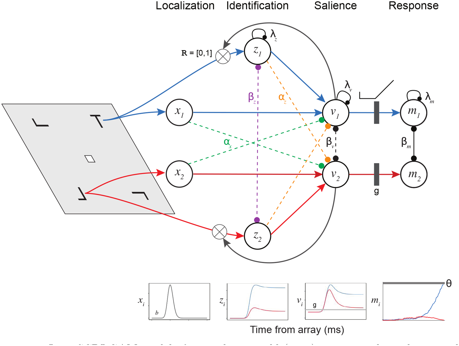
Joint SCRI-GAM model of Frontal Eye Field (FEF) neurons. The task is visual search, with a target “T” among a field of distractors shaped like rotated “L”s. An initial transient localization signal (xi) reflects the appearance of an object within a specific receptive field (RF) in a search display, and is equivalent for targets and distractors. The localization signal excites FEF visual neurons (vi) with the same RF and sends feedforward inhibition (αx) to FEF visual neurons centered on other RF’s. FEF visual neuron activation represents the momentary degree of salience attached to the part of the visual field that falls within their RF. FEF visual neurons receive a small amount of tonic excitation (b) and their spiking activity decays in the absence of additional excitation (λv). FEF visual neurons laterally inhibit one another (βv). FEF visual neurons can act as recurrent multiplicative gates (when ) to govern the rate at which a sustained identification signal (zi) grows toward an asymptotic value which tends to be higher for targets than distractors. These identification units are also subject to decay (λz) and laterally inhibit one another (βz). Identification units excite FEF visual neurons with the same RF and send feedforward inhibition (αz) to neurons with different RF’s. FEF visual neuron spiking activity that exceeds a threshold gate (g) excites FEF movement units mi with “movement fields” analogous to visual neurons’ RF’s. These movement units are subject to decay (λm) and laterally inhibit one another (βm). When a movement unit reaches a critical level of spiking activity (θ), a saccade is initiated to the unit’s movement field.