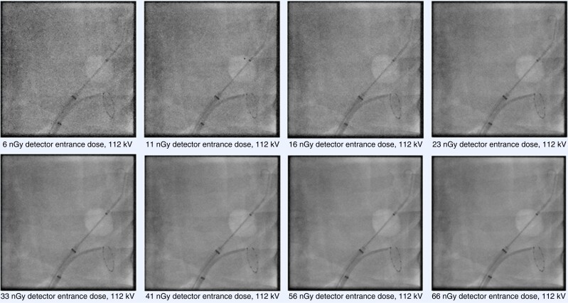
Figure 9.
Fluoroscopic acquisitions of electrophysiology catheters (cryoballoon and circular mapping catheter) placed inside a thoracic phantom model. The detector entrance doses of the fluoroscopy system (Artis zee, Siemens AG, Forchheim, Germany) were systematically increased from 6 nGy per pulse to 66 nGy per pulse at a constant X-ray tube voltage of 112 kV. Increasing the dose results in improvement of overall image quality, however relevant details (markers and electrodes) can still be identified at the lowest dose level (6 nGy).

