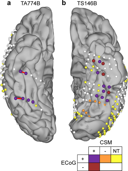
Figure 6. Cortical stimulation mapping (CSM) of reading.
Electrode localizations of the two patients who underwent reading CSM, highlighting electrodes active during electrocorticography (ECoG) of word reading (ECoG+; >20% BGA above baseline) and those leading to reading arrest during stimulation (CSM+). Sites not active during reading (ECoG-), not leading to reading disruption during CSM (CSM-) or not tested (NT) during CSM are noted. Red bars indicate the electrode pairs stimulated in Video 3. Patient TA774B was right hemisphere dominant for language, confirmed by intra-carotid sodium amobarbital injection.

