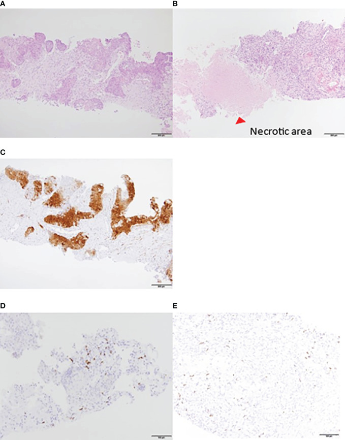
Figure 2.
Liver biopsy tissue. HE staining: increased necrotic area after treatment. (A) Pretreatment. (B) After two courses. IHC: P16 was positive. Local lymphocyte infiltration was observed before the initiation of treatment. (C) P16 was positive (D) CD8+ T lymphocytes pretreatment. (E) CD8+ T lymphocytes after two courses. Lymphocyte accumulation was observed around the liver. HE, hematoxylin and eosin; IHC, immunohistochemistry.

