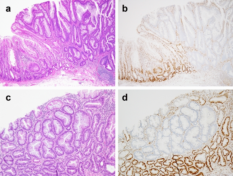
Fig. 2.
Case of sessile serrated lesion (SSL) with intestinal dysplasia (a, b), accompanied by minimal deviation dysplasia (c, d). a Non-dysplastic SSL (left) and intestinal dysplasia resembling the architecture of conventional adenoma (right). b Only intestinal dysplasia showed loss of MLH1 staining. (c, d) Minimal deviation dysplasia, showing large glandular structures, hypermucinous change, and loss of MLH1 staining, was incidentally identified apart from the intestinal dysplasia in location

