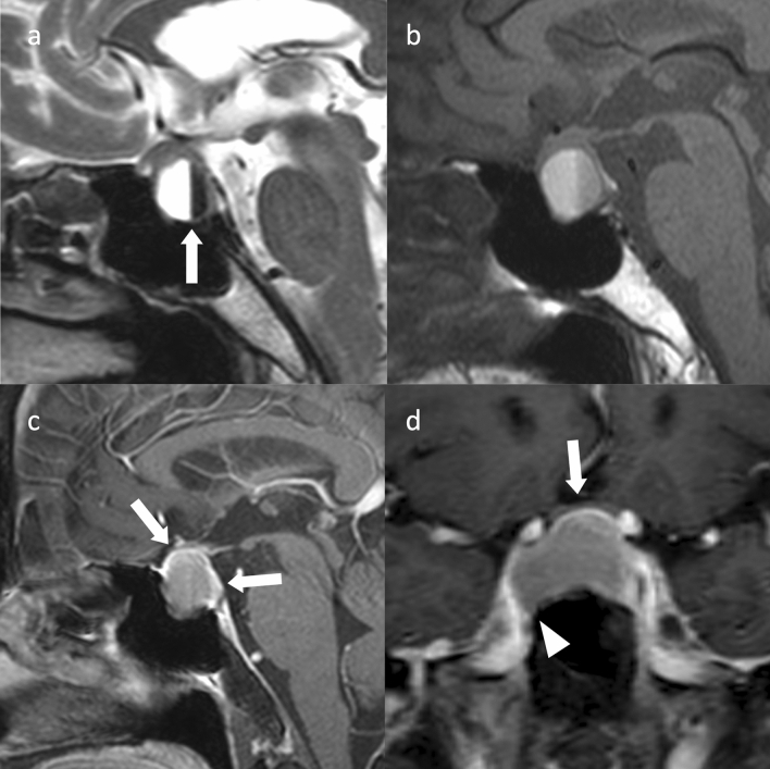
Fig. 5.
Cystic nonfunctioning PitNET/pituitary adenoma with internal bleeding. A female patient in her 70 s with bilateral auricular hemianopsia. A cystic mass is observed within the sella turcica to the suprasellar region. a Sagittal T2WI revealed a fluid–fluid level (arrow) with markedly hyperintensity anteriorly and markedly hypointensity posteriorly. b Sagittal T1WI shows hyperintensity anteriorly and mildly hypointensity posteriorly. c Sagittal contrast-enhanced T1WI shows a solid area of contrast at the limbus (arrow), which is believed to be a tumor component or pituitary gland. d The optic chiasm is slightly compressed upward by the mass on coronal contrast-enhanced T1WI (arrow). The mass protrudes into the right cavernous sinus and is classified as Knops grade 2 (arrowhead). The patient underwent surgery via the transsphenoidal sinus technique and diagnosed as a nonfunctioning PitNET/pituitary adenoma. No invasion of the cavernous sinus was observed

