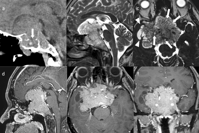
Fig. 7.
Silent corticotroph PitNET/pituitary adenoma. A male patient in his 40 s, with diplopia, disorientation, and cortisol levels within normal range. a Noncontrast CT shows destruction of the sella turcica (arrow) and invasion of the sphenoid sinus (asterisk) and the clivus (open circle). b Sagittal T2WI shows a mass with hyperintensity and iso-intensity areas. c Axial heavy T2WI shows small multiple cysts within the mass. Cysts around the mass likely represent trapped encysted pools of cerebrospinal fluid. d–f Contrast-enhanced T1WI shows a large mass centered in the sella turcica and invading the surrounding areas (the bilateral cavernous sinuses, and inferiorly, the sphenoid sinus and the clivus)

