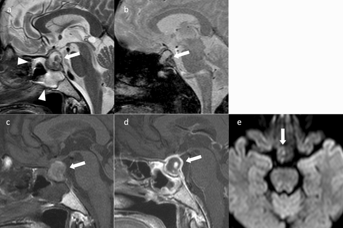
Fig. 8.
Pituitary apoplexy. A female patient in her 50 s, with headache, fever, and left ptosis. a Sagittal T2WI shows a mass in the sella turcica (arrow). The interior of the mass has a hyperintensity with some hypointensity regions. Mucosal thickening is observed in the sphenoid sinus (arrowhead). b Sagittal T2*WI reveals a hypointensity region in the mass, suggesting hemorrhage (arrow). c Sagittal T1WI shows a mildly hyperintensity mass (arrow). d Sagittal contrast-enhanced T1WI shows a ring-enhancing mass (arrow). e DWI shows iso-intensity (arrow). The patient underwent surgery via the transsphenoidal sinus approach, and hemorrhage and necrosis were observed

