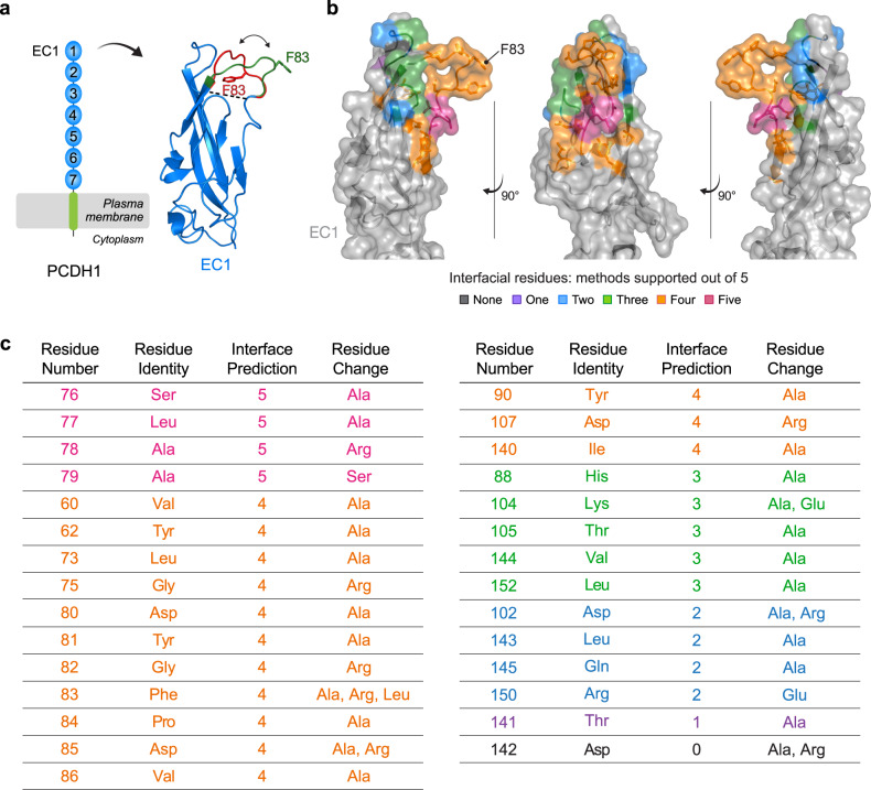
Fig. 3. Structure-based interfacial prediction reveals a surface patch on PCDH1 EC1 that potentially drives the interaction with ANDV and SNV Gn/Gc.
a Schematic representation of PCDH1 and crystal structure of EC1 (PDB 6MGA) displaying two modeled conformations (green: “open conformation”, red: “closed conformation”) for the disordered, uncrystallized loop comprising of residues 80–89. Residue F83 is indicated in each predicted loop conformation. b EC1 crystal structure in the “open conformation” displaying the EC1 residues chosen for mutational screening, ranked and colored according to the number of supporting algorithms. Structure adapted from PDB 6MGA. c List of the EC1 residues chosen for mutational screening in (b) ranked according to the number of supporting algorithms (interface prediction column). The amino acid substitution(s) are listed for each residue. (EC1, extracellular cadherin domain). Rankings of the residues in the PCDH1 ectodomain for each of the five complimentary algorithms can be found in Supplementary Data 2.

