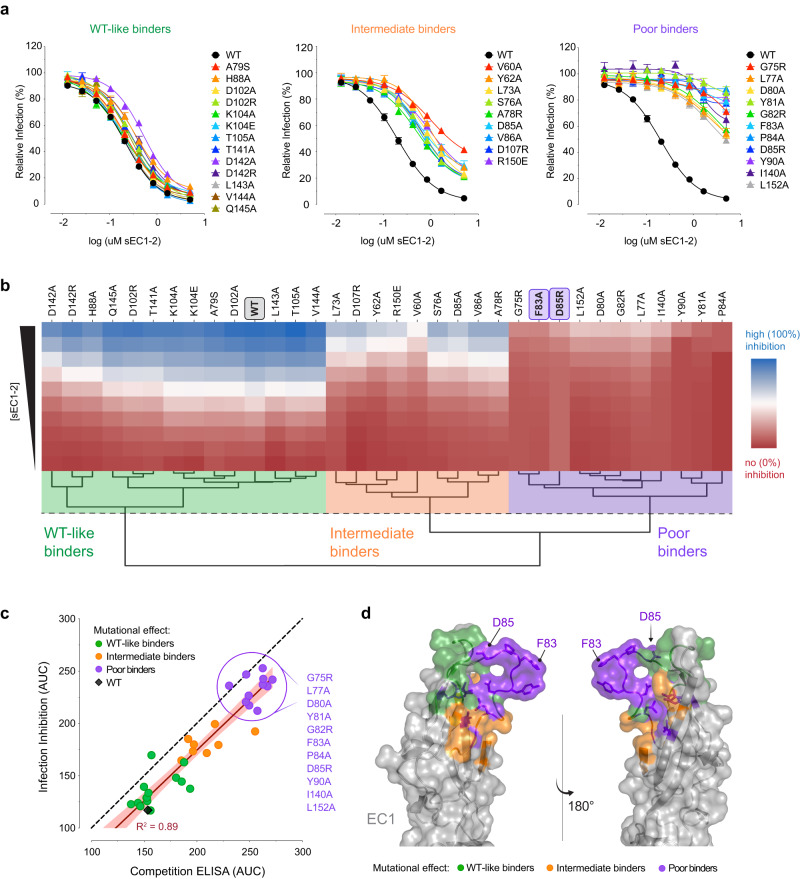
Fig. 5. Inhibition of ANDV Gn/Gc-mediated infection by mutant sEC1-2.
a WT and mutant sEC1-2 were tested on their ability to block rVSV-ANDV-Gn/Gc entry in primary human endothelial cells (HUVECs). The infectivity was normalized to that obtained without sEC1-2. Averages ± SEM: n = 6 wells of infected cells for each sEC1-2 dilution examined over three independent experiments [sEC1-2(T141A, L143A, V144A, Q145A, D85A, D142A, D142R, Y81A, F83A, D85A, I140A) have n = 7, sEC1-2(Y81A) has n = 6 for one dilution]. sEC1-2(WT) was used as a reference control and has n = 27 wells examined over 13 independent experiments. b Hierarchical clustering of WT and mutant sEC1-2 generated from sigmoidal curves of the infection-inhibition assay in (a). The dotted line denotes the height at which the dendrogram is cut to obtain three clusters representing varying degrees of inhibition of ANDV Gn/Gc-initiated infection; WT-like inhibition (green), intermediate inhibition (orange), and poor inhibition (purple). The red to blue colorbar ranges from 0 to 100 which is determined by the minimum and maximum values observed in the heatmap. c Area under the curve (AUC) for the binding activity of WT and mutant sEC1-2 to rVSV-ANDV-Gn/Gc, as determined by competition ELISA (see Fig. 4b), plotted against AUC values as determined by rVSV-ANDV-Gn/Gc infection-inhibition assay (a). The red line denotes the R squared value. A list of the sEC1-2 mutants that are classified as poor binders are listed to the right. d EC1 crystal structure in the “open conformation” displaying mutated residues representing three degrees of binding strength to ANDV Gn/Gc and inhibition of rVSV-ANDV-Gn/Gc infection. sEC1-2 mutants that bind and inhibit similarly to WT (WT-like binders), green; sEC1-2 mutants that display a mild reduction in binding and inhibition (intermediate binders), orange, and sEC1-2 mutants that display a strong reduction in binding and inhibition (poor binders), purple. Structure adapted from PDB 6MGA. Source data are provided as a Source Data file.

