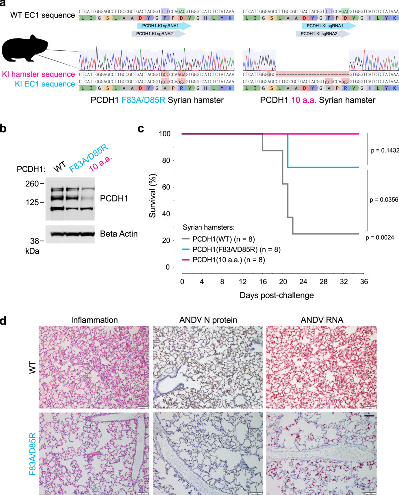
Fig. 8. Two point mutations in PCDH1 confer protection of Syrian hamsters against a lethal ANDV challenge.
a Reference nucleotide and amino acid sequence of PCDH1-EC1 Syrian hamster (WT, above) and representative sequences and trace files of Syrian hamsters after CRISPR-Cas9 genome editing [PCDH1(F83A/D85R), lower left] and [PCDH1(10a.a.) lower right]. The nucleotides encoding the corresponding human PCDH1-EC1 Gn/Gc-interacting residues, are highlighted: F83 in purple and D85 in green along with the location of the single guide RNAs (KI, knock-in; sgRNA, single guide RNA). b Immunoblot detecting PCDH1 in lung tissue lysates from WT or CRISPR knock-in mutant Syrian hamsters. Antibody targets PCDH1’s cytoplasmic tail. kDa, kilodalton. A representative blot from a single experiment of two independent experiments is shown. Uncropped blots in Source Data. c Syrian hamster ANDV challenge. Groups of WT, PCDH1(F83A/D85R), and PCDH1(10a.a.) CRISPR knock-in mutant hamsters were inoculated intranasally with ANDV (2,000 PFU). Mortality was monitored and hamsters were euthanized on day 35 post-exposure. One experiment was performed, with n = 8 hamsters for each group. Data was analyzed using two-sided, log-rank Mantel–Cox test. d Lung sections from WT and PCDH1(F83A/D85R) hamsters were collected 15 days post ANDV exposure. Representative histochemical images indicate inflammation in pulmonary tissue (left), ANDV nucleoprotein (N) (middle, tan staining), and ANDV RNA (right, red staining, detected by in situ hybridization). Representative images from one experiment from one out of three hamsters from each group are shown. Scale bars represent 100 µm. Figure (a) includes an image from Flaticon.com. Source data are provided as a Source Data file.

