
Figure 5.
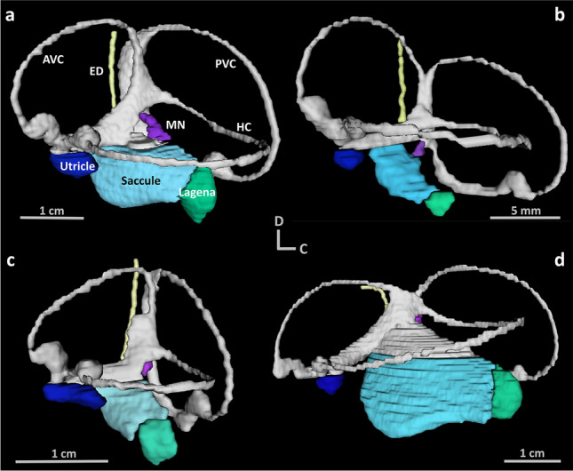
Three-dimensional visualisations.The inner ear from a (a) school shark (Galeorhinus galeus), (b) shortfin mako (Isurus oxyrinchus), (c) smooth hammerhead (Sphyrna zygaena), and (d) blue shark (Prionace glauca), illustrating the variation in different inner ear structures. AVC = anterior vertical canal, HC = horizontal canal, PVC = posterior vertical canal, MN = macula neglecta, ED = endolymphatic duct. All visualisations share the same orientation (D = dorsal, C = caudal).