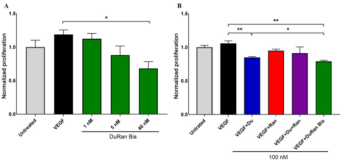
Figure 4.
Inhibitory effect of DuRan-Bis on the proliferation of U87MG cells, as shown in an XTT assay. (A) U87MG cells were treated for 48 h with 1 nM of VEGF either alone (black) or with 1 nM, 5 nM or 40 nM of DuRan-Bis (green). (B) U87MG cells were treated for 48 h with 1 nM of VEGF alone (black) or with 100 nM of Du (blue), Ran (red), a mixture of Du and Ran (purple), or DuRan-Bis (green). Cell viability measured using the XTT reagent was normalized to that of untreated cells (grey). Values are the means of triplicate experiments; bars represent SEM. *P < 0.05; **P < 0.01 (Student’s t-test, compared with cells treated with VEGF alone or with cells treated with VEGF and Du).

