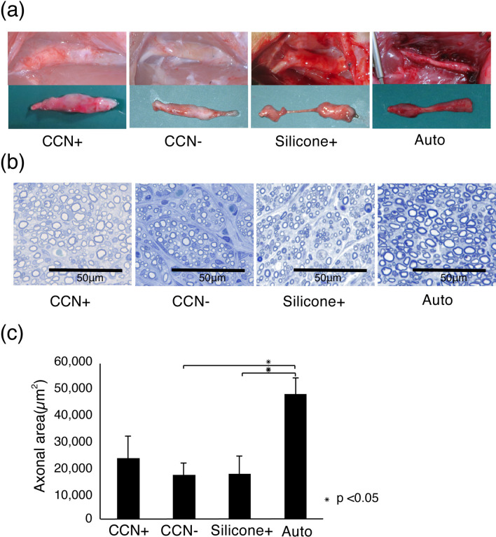
Figure 5.
Enhanced nerve regeneration by CCN with SCs. (a) Representative images of regenerated sciatic nerves 12 weeks after transplantation. The transplanted CCN in the CCN+ and CCN− groups remains, despite the occurrence of bio-absorption. (b) Toluidine blue staining in the central axial sections reveals regenerated nerve fiber in all groups. (c) Quantitative analysis of the axonal area of the regenerated nerve fibers (axonal area = cross-sectional area × axon density). The CCN+ group has higher axonal area value than the CCN− and silicone+ groups, but the difference among the three groups is not significant.

