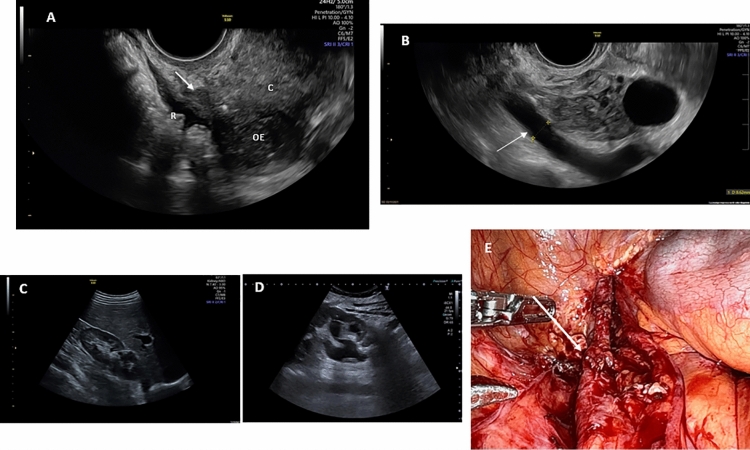
Figure 1.
(A) Arrow: Sonographic image of hypoechoic thickening of the uterosacral ligament; C: Cervix; R: Rectum; OE: Ovarian Endometrioma. (B) Arrow: Sonographic image of ureteral dilatation by the left adnexal space. (C) Normal kidney. (D) Hydronephrosis secondary to distal ureteral stenosis due to deep endometriosis nodule. (E) Surgical image of distal ureteral stenosis (arrow) and upper dilatation due to deep endometriosis pelvic nodule.

