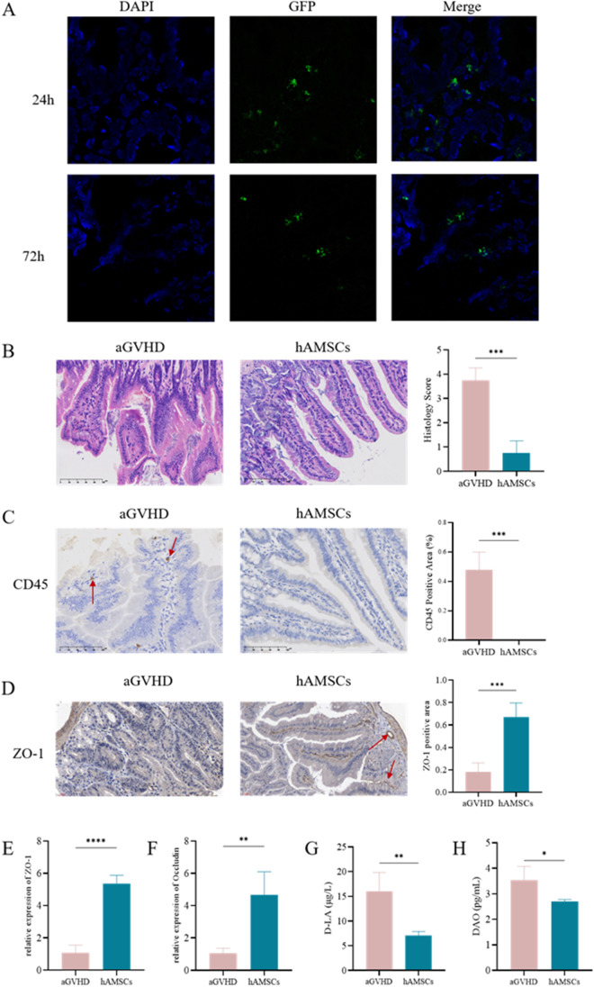
Fig. 5.
hAMSCs ameliorated intestinal barrier dysfunction in aGVHD mice. (A) GFP-labeled hAMSCs infiltrated into the intestines. (B) Representative microscopic pictures of H&E staining of intestines (400 ×) and histology score (based on the tissues structure destruction and lymphocyte infiltration) (n = 4). (C) Representative microscopic pictures of immunohistochemistry (400 ×) of intestines and the proportion of CD45 positive area (n = 4). (D) Representative microscopic pictures of immunohistochemistry (400 ×) of intestines and the proportion of ZO-1 positive area (n = 4). (E) mRNA expression levels of ZO-1 and Occludin (n = 3). (F) Plasma levels of D-LA and DAO (n = 4). Values were presented as mean ± SD

