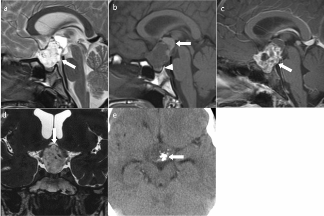
Fig. 1.
Adamantinomatous craniopharyngioma. A 13-year-old girl with headache and vision loss. a Sagittal T2WI shows a multifocal cystic mass above the sella turcica (arrow). The pituitary gland is observed within the sellar turcica. b Sagittal T1WI shows some cysts with mild hyperintensity (arrow) compared to white matter. Normal hyperintensity in the posterior pituitary gland is observed (arrowhead). c Sagittal contrast-enhanced T1WI shows heterogeneous contrast enhancement. The pituitary stalk is inside the mass and cannot be identified (arrow). d Coronal heavy T2WI shows the optic chiasm compressed by the mass (arrow). e Non-contrast computed tomography shows calcification inside the mass (arrow)

