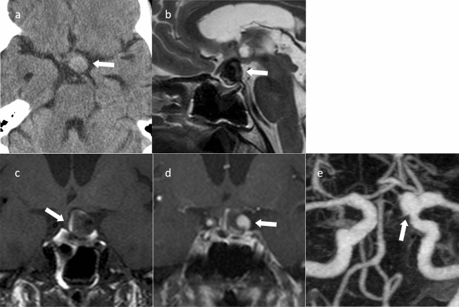
Fig. 13.
Internal carotid-posterior communicating artery aneurysm with partial thrombosis. An approximately 70-year-old female with headache. a Computed tomography shows a high-density mass in the suprasellar region (arrow). b Sagittal T2WI shows a hypointensity mass above the sella turcica (arrow). c Coronal T1WI shows hypointensity in the mass but with a hyperintense area at the limbus, indicating a thrombus (arrow). d Coronal contrast-enhanced T1WI shows a nodular area with contrast enhancement in the mass (arrow). e Magnetic resonance angiography shows an aneurysm in the posterior communicating artery of the right internal carotid artery (arrow)

