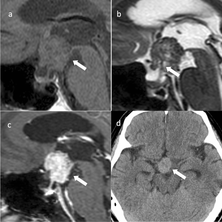
Fig. 2.
Papillary craniopharyngioma. An approximately 50-year-old male with eye floaters. a Sagittal T1WI shows a mass with isointensity compared to white matter from the suprasellar region into the third ventricle (arrow). b Sagittal T2WI shows some small cyst-like hyperintensity areas in a iso- to hypointense mass compared to white matter (arrow). A duct-like recess is observed at the base of the mass contiguous with the pituitary stalk (arrow). c Sagittal contrast-enhanced T1WI shows almost uniform and strong contrast enhancement (arrow). d Non-contrast computed tomography shows mild high density (arrow). The patient underwent surgery via nasal endoscopy, and the mass was confirmed as a papillary craniopharyngioma arising from the base of the third ventricle

