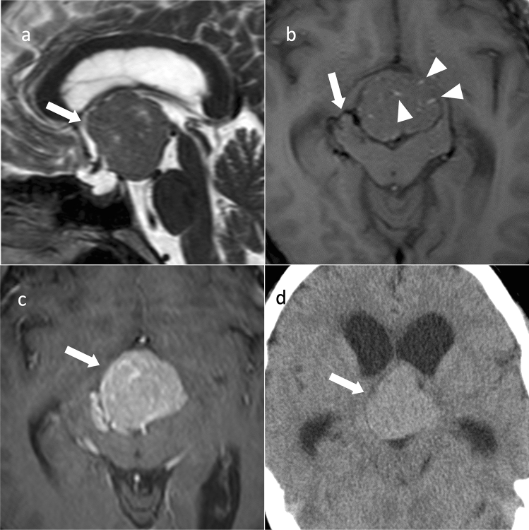
Fig. 3.
Pituicytoma. An approximately 40-year-old male with mild cognitive decline. a Sagittal T2WI shows a lobulated mass with isointensity compared to gray matter in the suprasellar region (arrow). b Axial T1WI shows a flow void around the mass (arrow). Small hyperintense areas within the mass show slow-flowing vessels (arrowheads). c Contrast-enhanced T1WI shows strong and uniform contrast enhancement (arrow). d Non-contrast CT shows mild high density (arrow). Ventricular enlargement is observed

