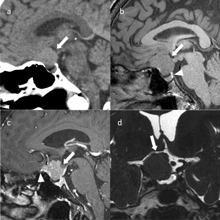
Fig. 5.
Tuberculum sellae meningioma (meningothelial meningioma). An approximately 60-year-old female with decreased right visual acuity. a Sagittal non-contrast computed tomography showing a mildly hyperdensity mass compared to white matter in the suprasellar region (arrow). No obvious thickening of the bone adjacent to the mass is observed. b On sagittal T1WI, the mass shows isointensity compared to gray matter (arrow). Normal posterior pituitary gland hyperintensities are observed (arrowhead). c Sagittal contrast T1WI shows uniform and strong contrast enhancement (arrow). The dural tail sign is observed anterior to the mass (arrowhead). d Coronal heavy T2WI shows compression of the right optic nerve by the mass (arrow). Craniotomy was performed, and the tumor was confirmed as a meningothelial meningioma

