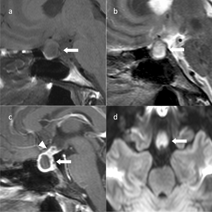
Fig. 8.
Pituitary abscess. An approximately 20-year-old female with headaches and nausea. Sagittal T1WI (a) and sagittal T2WI (b) show a cystic mass within the sellar turcica to the suprasellar region (arrow). Sagittal T2WI (b) failed to show hypointense regions of the peripituitary region (parasellar T2-dark sign). c Sagittal contrast-enhanced T1WI shows contrast enhancement of the cyst wall (arrow) with an enlarged pituitary stalk (arrowhead). d Diffusion-weighted imaging (DWI) shows marked hyperintensity inside (mean apparent diffusion coefficient = 0.5 × 10⁻³ mm2/s) (arrow)

