Fig. 9.
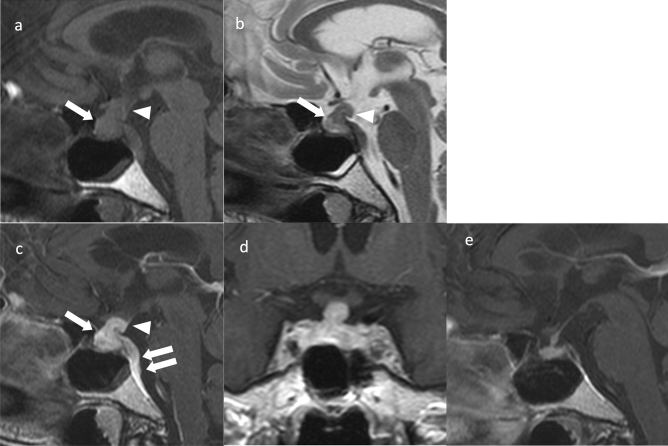
Lymphocytic hypophysitis. An approximately 80-year-old female with left oculomotor nerve palsy. Sagittal T1WI (a) and sagittal T2WI (b) show an enlarged pituitary gland (arrows) and pituitary stalk (arrowheads). Sagittal T1WI (a) shows no hyperintensity in the posterior pituitary. c Sagittal contrast T1WI shows comparative homogeneous contrast effects of the pituitary gland (arrow) and stalk (arrowhead). A thickened dura mater is observed on the dorsal surface of the clivus (double arrow). d Coronal contrast-enhanced T1WI shows a symmetrical enlarged pituitary gland and stalk. e Sagittal contrast-enhanced T1WI after 3 months of oral steroid treatment shows a reduction in the size of the pituitary gland and stalk. Dural thickening also improved

