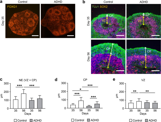
Fig. 1.
Comparison of layer structures in telencephalon organoids derived from ADHD and control. (a) Expression of FOXG1, a telencephalon-specific marker protein, at day 35 of differentiation. Scale bars: 200 μm. (b) Layer structures in the organoids at day 35 and 56. Both control-derived and ADHD-derived organoids showed SOX2-expressing neural stem cells in ventricular zone-like structures. The neuron-specific marker protein TUJ1 was expressed in the outer cortical plate–like structure. Scale bars: 40 μm. (c) Thickness of neuroepithelium-like structures (NE). (d) Thickness of cortical plate–like structures (CP). (e) Thickness of ventricular zone–like structures (VZ). Control: day 35 (n = 30), day 56 (n = 33); ADHD: day 35 (n = 30), day 56 (n = 41). *P < 0.05, **P < 0.01, ***P < 0.001

