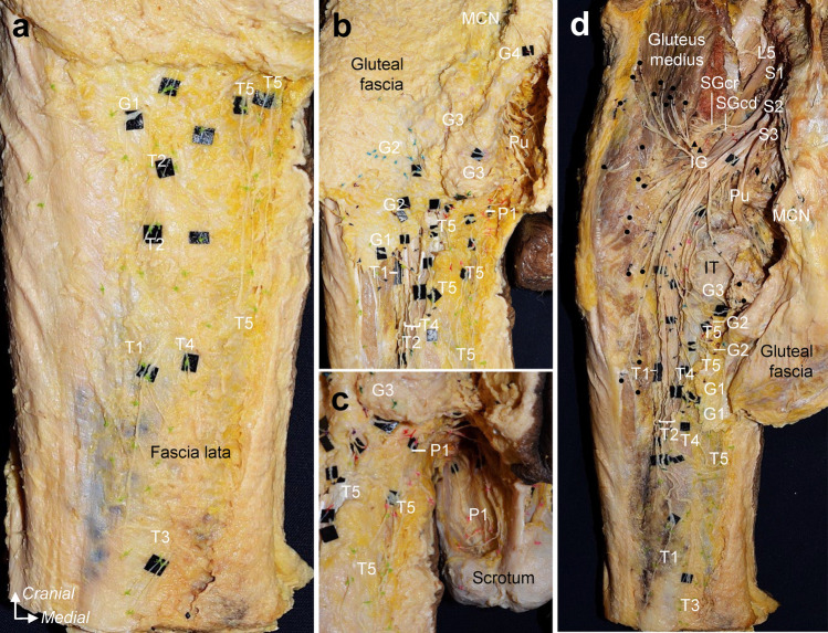
Fig. 1.
Dissection of the cutaneous branches to the posterior thigh, inferior gluteal, and perineal regions (Specimen no. 3; left, male). a: Posterior thigh region. After the removal of the skin, the thigh branches subsequent to penetrating the fascia lata were recorded with colored strings. b: Inferior gluteal region. The facia lata was partly cut and removed, preserving the area where the cutaneous nerve penetrates the facia lata as much as possible, and the gluteal branches were recorded with colored strings before removing the gluteal fascia and gluteal maximum. c: Perineal region. Perineal branches were also recorded with colored strings. d: After recording the point at which each cutaneous nerve penetrated the fascia lata and ran the course concerning the ischial tuberosity and inferior margin of the gluteus maximus, we removed the fascia lata, gluteus maximus, sacrotuberous ligament, and bony elements of the sacrum and ilium to analyze the origin of the cutaneous nerves. T, G, and P indicate the thigh, gluteal, and perineal cutaneous branches, respectively, and are numbered from the lateral side. Circle = nerve to the gluteus maximus; Diamond, nerve to coccygeus and levator ani; IG, inferior gluteal nerve; IT, ischial tuberosity; MCN, medial cranial nerve; Pu, pudendal nerve; SGcd, caudal part of the superior gluteal nerve; SGcr, cranial part of the superior gluteal nerve; Ti, tibial nerve; triangle, nerve to piriformis

