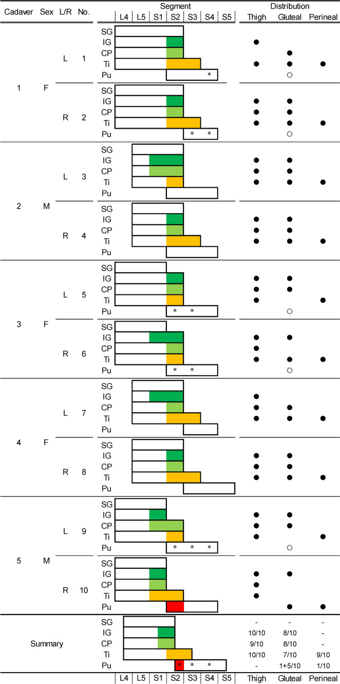
Fig. 2.
Relationships between the origin and distribution of the posterior femoral cutaneous nerve. The figure shows the segmental arrangement of the main nerves of the sacral and pudendal plexuses and their relation to the posterior femoral cutaneous nerve in all specimens. Colored areas (green, inferior gluteal nerve; light green, common peroneal nerve; orange, tibial nerve; and red, pudendal nerve) indicate the origin and segment of the posterior femoral cutaneous nerve. The black circle indicates the presence of the posterior femoral cutaneous nerve distributed in the thigh, gluteal, and perineal regions. Asterisks and white circles indicate the presence and segment of the cutaneous branches originating from the pudendal nerve to the gluteal region. CP, common peroneal nerve; F, female; IG, inferior gluteal nerve; L/R, left or right sides; M, male; Pu, pudendal nerve; Ti = Tibial nerve.

