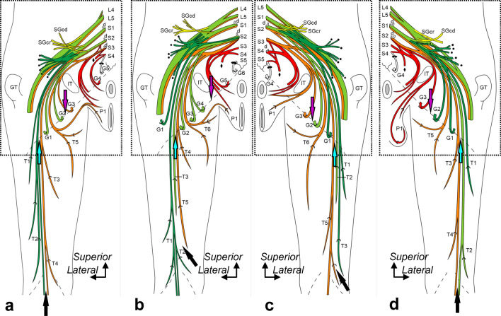
Fig. 3.
Origin, course, and distribution of the posterior femoral cutaneous nerve. Schematic illustrations of the buttocks and posterior thighs. a: Specimen No. 1 (left, female), b: Specimen No. 5 (left, female), c: Specimen No. 8 (right, female), and d: Specimen No. 10 (right, male). The thigh, gluteal, and perineal cutaneous branches are indicated by T, G, and P, respectively, numbered from the lateral side. The dotted oblique line indicates the inferior margin of the gluteus maximus, and the recurrent course of the distal cutaneous branches is shown in Fig. 4 (indicated by the boxed region). The posterior femoral cutaneous nerve originates from the following nerves comprising the sacral and pudendal plexus: inferior gluteal (green), common peroneal (light green), tibial (orange), and pudendal nerves (red). Based on the stratification relationship among these nerves, the blue and purple arrows at the inferior margin of the gluteus maximus indicate the dorsoventral boundaries of the thigh and gluteal cutaneous branches, respectively. The black arrow at the distal posterior thigh indicates the dorsoventral boundary of the thigh. Circle, nerve to the gluteus maximus; d, dorsal rami; Diamond, nerve to coccygeus and levator ani; GT, greater trochanter; L, (e.g., L4) the lumbar nerve; IT, ischial tuberosity; S, (e.g., S1) the sacral nerve; SGcd, caudal part of the superior gluteal nerve; SGcr, cranial part of the superior gluteal nerve; and triangle, nerve to the piriformis.

