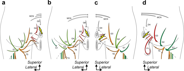
Fig. 4.
Posterior femoral cutaneous nerve distribution in the gluteal region. Schematic illustrations of the buttocks. a: Specimen No. 1 (left, female), b: Specimen No. 5 (left, female), c: Specimen No. 8 (right, female), and d: Specimen No. 10 (right, male). The corresponding illustrations deep to the gluteus maximus of a–d are shown in the boxed region of Fig. 3a–d. The thigh, gluteal, and perineal cutaneous branches are indicated by T, G, and P, respectively, numbered from the lateral side. The cutaneous branches are colored based on their origin, such as the inferior gluteal (green), common peroneal (light green), tibial (orange), and pudendal nerves (red). The yellow arrow indicates the boundary between the ventral and dorsal rami. Dotted oblique line = inferior margin of the gluteus maximus, and MCN = medial cluneal nerve.

