
FIGURE 3.
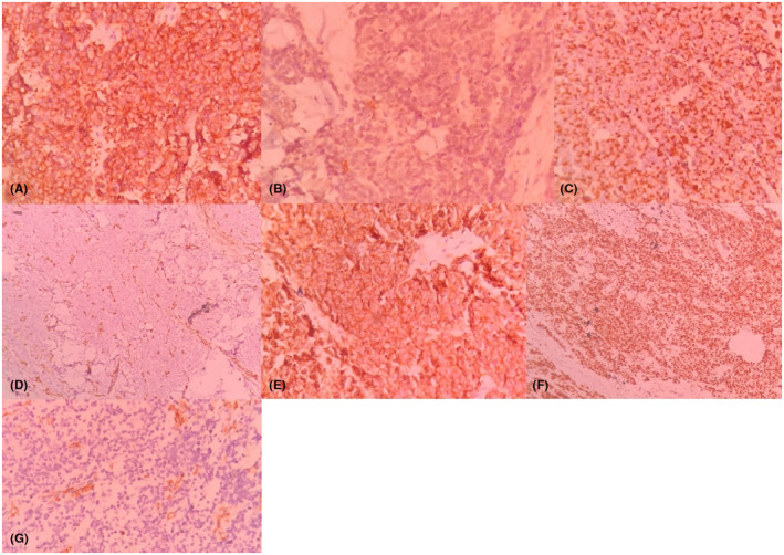
Immunohistochemical staining, the tumoral cells are positive for synaptophysin, (40×) (A), focally positive for S‐100 (40×) (B), positive for AE1/AE3 (40×) (C), negative for Vimentin (10×) (D), positive for CD56 (40×) (E), positive for Ki67 (10×) (F), and negative for CD45 (40×) (G).