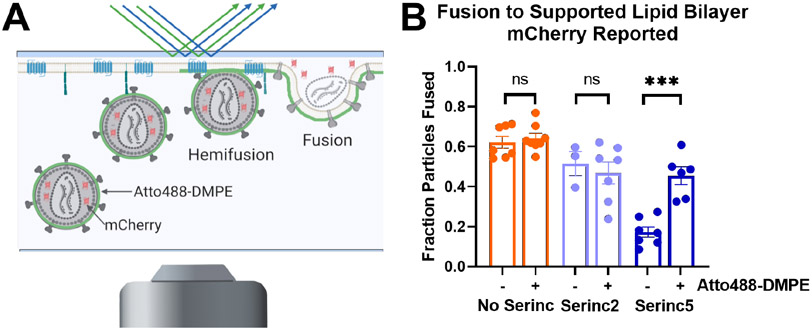
Figure. 3. Lipid composition of the viral membrane affects Serinc restriction of HIV membrane fusion.
(A) Diagram of TIRF microscopy-based single HIV pseudovirus particle fusion assay. Pseudovirus binding to a supported planar plasma membrane containing CD4 receptor and CCR5 co-receptor is reported as a sudden appearance of bright puncta that remain stationary for several imaging frames. Fusion is reported by a decrease in fluorescence over several imaging frames as a genetically encoded content marker, mCherry, diffuses away. (B) Incorporation of Atto488-DMPE increases fusion of HIV pseudovirus particles containing Serinc5 but does not affect fusion of particles without Serinc or containing Serinc2. Each data point represents the fraction of particles fused on a separately prepared bilayer. Reproduced from ref. 3, copyright year 2020. ***, p < 0.001; ns, not significant by multiple unpaired t-tests via the Holm-Sidak method. Each condition includes data from at least three distinct preparations of pseudovirus.

