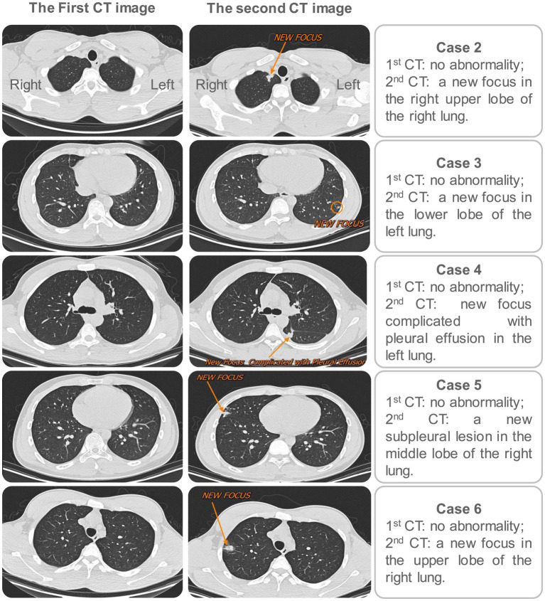
Figure 3.
Comparison of the first and second screenings of chest CT for the five secondary cases. In the first screening, no abnormalities were found in the chest CT for the 5 cases. Three months later, the second screening of chest CT was performed, and new lesions were observed in the lungs of all five cases. Left: the first CT image for each case; Right: the second CT image for each case.

